


What is Included in a Comprehensive Eye Examination?
During the exam, the medical team thoroughly analyzes all the structures of the eye:
- Refraction
- Eyelashes
- Eyelids
- Tear FILM
- Cornea, iris and lens
- Intraocular pressure (IOP) and the optic nerve
- Retina
- Macula
- Eye movements and pupillary reflexes


What are cataracts?
It is the loss of transparency of the lens that produces a progressive loss of vision.
Symptoms
- Blurry vision
- Loss of color sharpness
- Poor night vision
- Frequent glares
- Foggy vision

Normal eye

Eye with cataract

Normal vision

Vision with cataract
What is the treatment?
The only effective way to remove a cataract is through surgery . You don’t have to wait until you lose all your vision to have surgery. In fact, early cataracts require simpler surgery with faster visual recovery. It is a surgery that offers definitive results.
What does the surgery involve?
The operation consists of removing the opacified lens and placing an intraocular lens in its place , which may be monofocal (to correct vision at any distance) or multifocal (to correct vision at any distance).
Surgical techniques
The most advanced technique today is called phacoemulsification , which aims to replace the opaque crystalline lens with an artificial lens that allows the patient to recover lost vision.
What type of lens can we implant?
Monofocal lens
We replace the opaque lens with an artificial lens, with the necessary prescription so that the eye recovers vision for far.
Multifocal lens
We implant a lens that will allow the patient to see both far and near , in most cases, without needing to use glasses.
Toric lens
It is a lens that can be either monofocal or multifocal and also corrects astigmatism.
Advantatges of cataract surgery:
- Safe and effective technique
- Quick and painless intervention
- Outpatient surgery
- With topical anesthesia
- No waiting list
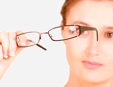
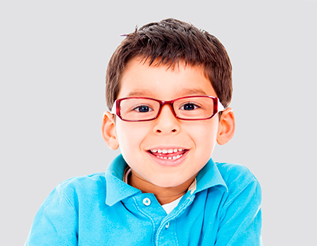
What are refractive errors (myopia, hyperopia and astigmatism)?
In order to see images clearly, they must be focused exactly on the retina. An eye is said to have a refractive error when the power the lenses (crystalline lens and cornea) focuses the images either in front (myopia) or behind (hyperopia) of the retina.
Myopia
A person with myopia has difficulty (blurred vision) focusing on distant objects, while seeing nearby objects correctly. Myopia can cause headaches or eye fatigue.
Farsightedness
Farsighted people have a blurred perception of nearby objects , unlike nearsighted people. Farsightedness can cause headaches, tearing, or frequent blinking.
Astigmatism
A person with astigmatism perceives things as distorted , both near and far, due to a problem with the curvature of the cornea. Astigmatism is often associated with myopia or hyperopia.
All these refractive defects can be corrected with glasses, contact lenses or permanently through refractive surgery.
What is laser refractive surgery?
It is an effective and safe technique for correcting myopia, hyperopia and astigmatism.
By applying the laser, the curvature of the cornea is modified, which allows the refractive defect to be corrected precisely, with minimal discomfort for the patient and with a rapid recovery of their visual function.
What techniques exist?
Among the different existing techniques, it is worth highlighting the Lasik, PRK and, in cases of high myopia, phakic lenses (ICL).
After a personal visit and a thorough examination, the doctor decides the most appropriate technique for each patient.
Advantages of refractive surgery:
- Safe and effective technique
- Thousands of people operated on
- Quick and painless intervention
- Outpatient surgery
- With topical anestesia
- No waiting list
What is presbyopia?
It is a progressive difficulty in reading or focusing on nearby objects . It occurs physiologically in all people from the age of 40-45 and affects 100% of the population over 50 years of age.
Why does it appear?
As a result of aging:
The ciliary muscle loses elasticity and power.
The lens loses elasticity.
The eye loses its ability to focus on objects up close.
The person begins to move away from the text , looking for an ideal focusing distance or looking for well-lit areas.
Who does it affect?
Presbyopia is inevitable. It affects both patients who have never worn glasses and those who have some refractive defect (myopia, hyperopia or astigmatism).
Other factors that may worsen presbyopia are:
- Diabetes
- Anemia
- Certain medications
Symptoms
- Difficulty reading or focusing on nearby objects.
- Increasing the distance between the object and the eyes, a characteristic sign of presbyopia (extending the arms to read).
- Need for more light for reading.
- Headache, feeling of heaviness in the eyes, itchy and red eyes.
How can it be treated?
Traditional correction is done with progressive glasses.
Nowadays, it can be permanently corrected by surgery, which will allow, in most cases, to see both near and far without needing to use glasses.
Surgery may involve implanting an intraocular lens.
Advantages of cataract surgery:
- Safe and effective technique
- Quick and painless intervention
- Outpatient surgery
- No waiting list
What is glaucoma?
This pathology is caused by damage to the optic nerve, usually caused by an increase in eye pressure. Glaucoma leads to irreparable damage to the optic nerve, which causes a progressive loss of vision that can even lead to total blindness.
Symptoms
- Frequent change of glasses.
- Difficulty seeing in dark places.
- Loss of peripheral vision.
These symptoms are not always signs of glaucoma. By the time a patient begins to notice visual impairment, his or her vision may already have been significantly affected. For this reason, glaucoma is known as the silent eye disease , since it does not give any warning (the symptoms do not cause pain or discomfort).
Treatment
The goal of treatment is to preserve vision by reducing intraocular pressure.
In the initial treatment of the disease, eye drops are usually used to reduce the pressure in the eye, but in some cases the reduction in pressure may be insufficient and surgical treatment may be necessary.
How to prevent glaucoma
The glaucoma screening test is quick and painless. Everyone over 35 years of age should have their intraocular pressure (IOP) measured at least once a year and have an eye examination to assess the state of the optic nerve.
If the pressure is borderline or the optic nerve looks suspicious of glaucoma, specific tests will be performed and the person will be monitored more frequently.
About glaucoma:
- Glaucoma is known as the silent eye disease.
- It is very important to monitor intraocular pressure (IOP).
- Periodic eye examination.
What is the retina?
It is the light-sensitive layer of tissue located at the back of the eyeball. Images passing through the lens of the eye are focused on the retina, which converts these images into electrical signals and sends them along the optic nerve to the brain.
Retinal pathologies:
- AMD (Age-Related Macular Degeneration)
- Diabetic Retinopathy
- Retinal detachment
- Vitreous detachment
- Hypertensive retinopathy
- Epiretinal membrane
- Macular hole
- Retinitis pigmentosa
What is AMD?
It is a degenerative disease of the macula or central area of the retina. It is one of the most common causes of vision loss in people over 65 years of age.
Symptoms
- Difficulty reading
- Blurred central vision
- Black spot in the center of the vision and distorted lines
- Color alteration or deformed straight lines
Type of AMD
- Dry AMD, in which the tissue in the macular area gets thin.
- Wet or atrophic AMD is usually more aggressive and progresses more quickly.
Treatment
- There is currently no treatment for dry AMD.
- The usual treatment for wet AMD is usually with drugs that are administered through intravitreal injections.
When to see an ophthalmologist?
- Injections to treat AMD
- If you “see floaters” visit urgently
- If you are diabetic, do at least one a year
What is blepharoplasty surgery?
This is surgery that corrects changes in the eyelids caused by aging. This surgery removes excess skin, muscle and fat, which cause a more tired and sad look, as well as a feeling of heaviness and even difficulty opening the eyes.
Surgery is usually performed under local anesthesia with sedation and without hospitalization. It is possible to perform personalized surgery to correct changes caused by the passage of time, without changing the usual features and with minimal risks.
Type of blepharoplasty:
- Upper blepharoplasty: The goal is to remove excess skin from the upper eyelids and clear the look.
- Lower blepharoplasty: The goal is to remove bags under the lower eyelids. The skin can also be tightened when necessary. It can be performed using a traditional technique or via a transconjunctival approach.
Is it a safe intervention?
Blepharoplasty is the most common eyelid operation and is completely safe. The surgeon seeks to improve the patient’s aesthetics, but always prioritising the function of the eye and its attached facial structures. In fact, problems with excess skin on the eyelids can also interfere with the patient’s field of vision, which is restored after the operation.
What are the advantages of going to an ophthalmology specialist for blepharoplasty?
The ophthalmologist specializing in Ocular Plastic Surgery knows the anatomy and physiology of both the eye and the eyelids, and is trained in minimally invasive microsurgery, so he can achieve a good aesthetic result with minimal risks, without altering the important protective function of the eyeball, achieving great harmony between aesthetics and functionality.
Oculis has an Ocular Plastic Surgery Unit with ophthalmologists specialized in eyelid surgery.
About blepharoplasty:
- Eliminates the tired look effect
- Corrects drooping eyelids
- Eliminates bags under the eyes
- Rejuvenates
- Opens the field of vision
What is the cornea?
The cornea is the structure of the eye that allows light to pass from the outside to the inside of the eye and protects the iris and the lens. To ensure its function, it must be transparent and maintain an appropriate curvature.
It has two main functions:
- Acts as a protective shield
- Controls the focus and entry of light into the eye
Corneal diseases
Various corneal diseases can alter its characteristics and cause impaired vision.
- Conjunctivitis
- Dry eye
- Pterygium
- Keratoconus
- Keratitis
- Corneal dystrophies
- Herpes
- Corneal opacity
Sometimes, refractive errors (myopia, hyperopia and astigmatism) also originate in the cornea.
Dry eye
Dry eye is a very common pathology that represents 30% of all ophthalmology consultations.
It is more common after the age of 40. It occurs as a result of the alteration of the tear film that covers the eye and damages its external surface.
Some of the discomforts it causes are:
- Itching sensation
- Stinging sensation
- Sensation of having sand in the eye
- Difficulty opening eyes in the morning
- Slight sensation of blurred vision
Conjunctivitis
Conjunctivitis is an infectious process that occurs in the conjunctiva. It can spread and also affect the cornea, compromising vision. It is more common in spring and autumn.
Symptomatic differences between conjunctivitis and dry eye
- Conjunctivitis: more intense redness and tearing
- Dry eye: no tearing and difficulty opening the eye
What are tear ducts?
The tear duct is the natural route for the evacuation of tears from the eye to the nasal passages. Obstruction of this duct is the most common cause of tearing and eye infections.
The blocked tear duct
A blocked tear duct is a condition that is caused by inflammation in the tear sac and the tear duct.
When this duct is blocked, the tears have nowhere to go and overflow, causing constant tearing, irritation and infection.
Type of blocked tear duct
- Acquired: This is the most common type. It occurs when the blocked tear duct develops in adults or children. It mainly affects women.
- Congenital: There is a congenital form that affects newborns, and which usually resolves on its own in 90% of cases before the age of one.
Causes
Blockages are caused by infections, foreign bodies or other causes, such as:
- Nasal trauma
- Neoplasms of the tear sac or duct
- Bone abnormalities
- Inflammatory diseases such as sarcoidosis or Wegener’s disease
- As a result of surgical trauma
Treatments
Treatment is always surgical and will depend on the cause of the obstruction.
What is the surgery like?
One of the surgeries for obstructed tear ducts is balloon catheter acrioplasty . It is a minimally invasive technique, with no scars, minimal post-surgical complications, rapid recovery and a very high success rate. Dacryocystorhinostomy can also be performed for low-level obstruction and Lester Jones tube placement for upper tear duct obstruction.
About the tear ducts:
- Epiphora (constant and excessive tearing)
- Chronic conjunctivitis and eczema on the eyelids
- There may be swelling and pain in the area of the tear sac.
What is strabismus?
Strabismus is the misalignment of the eyes.
If the muscles of one eye do not work in a coordinated manner with those of the other eye, strabismus occurs, that is, a deviation of the eyes commonly known as “crossed eye.”
It produces a loss of stereoscopic vision (depth) that may be reversible depending on the age of the patient and the type of strabismus, and can produce amblyopia of the deviated eye (lazy eye) that may require treatment with optical correction and patching.
Type of strabismus
Convergent strabismus: This is the most common type of strabismus, when the eyes deviate towards the nose. It is called esotropia. It can be corrected with optical correction (glasses), surgery or a combination of both. The so-called accommodative esotropia is treated with glasses and surgery is not necessary. However, there are other strabismus that improve with glasses, but are not completely resolved, so surgery and glasses will be necessary (partially accommodative esotropia). The third group, non-accommodative esotropia, cannot be corrected with glasses and requires surgery.
Divergent strabismus: These are eyes that deviate outwards. They are the second most common. They tend to become unbalanced in situations of looking far away, tiredness, stress, when the person distracted or bothered by bright light. They can improve with optical correction but most require surgical intervention. They are called exotropias.
Vertical strabismus: The eye deviates upwards or downwards. These are called hypertropia or hypotropia respectively. They usually require surgical correction.
Congenital strabismus: Present at birth and not usually improved with glasses, they require surgery in the vast majority of cases.

Normal eyes

Convergent strabismus

Divergent strabismus

Vertical strabismus
Treatments for strabismus
- Surgery: Surgery involves shortening, lengthening or modifying the position of the muscles to restore parallelism of the eyes. Is common and for this reason, in a high percentage of patients, reoperation is necessary after months/years.
- Glasses: These affect the position by changing the person’s reaction to focusing. Prisms change the direction of light and therefore the images, which causes the position of the eye to change.
- Occlusions: Ocular occlusions (patches) do not serve to correct strabismus but they do serve to prevent or treat an amblyopic (lazy) eye and are a very important treatment in the overall management of a child with strabismus.
- Visual therapy: In certain cases it can be corrected with eye exercises.
